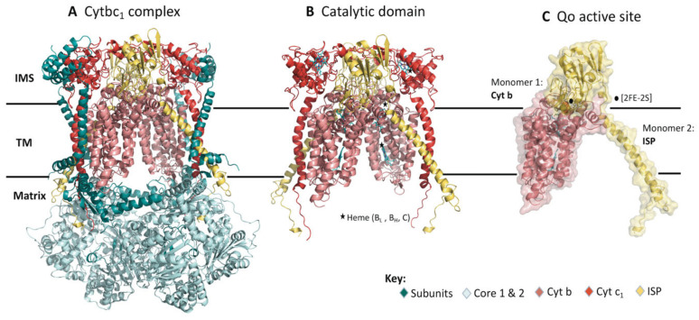
Figure 1.
The structural model of the dimeric bc1 complex in bovine (PDB ID: 1PP9). (A) The crystal structure in cartoon representation showing the multi-subunit dimer. The different subunits are color coded (key provided) and labeled accordingly. (B) Catalytic domain composed of Cytb, cyt c1 and ISP spans across the inner mitochondrial membrane. (C) Heterodimeric structure made up of Cytb and ISP both of which form the Qo active site. Black stars and circles represent the hemes and [2FE-2S] cluster, respectively. (IMS—Intermembrane space, TM—transmembrane).

