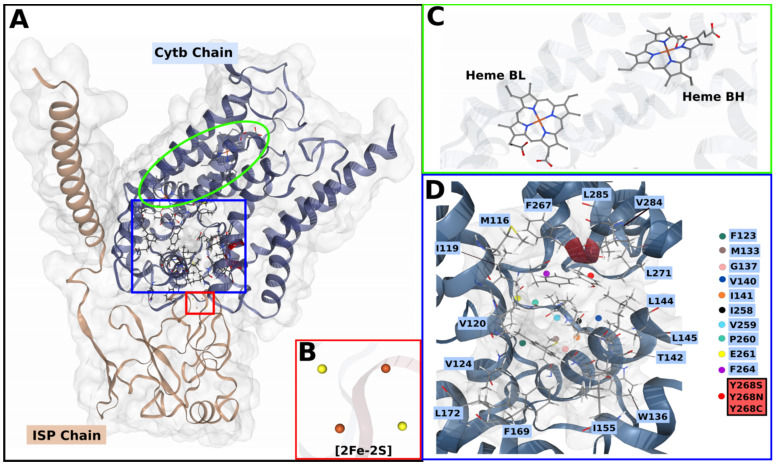
Figure 2.
3D cartoon representation of PfCytb-ISP protein complex. (A) PfCytb-ISP structure with different subunits color coded (key provided) and labelled accordingly. The red, green and blue delimitations indicate the [2FE-2S] cluster, the heme groups and the active site, respectively. (B) [2FE-2S] cluster, showing sulphur atoms in yellow and Fe2+ atoms in orange. (C) Heme bL and bH groups. (D) The zoomed in active site showing its contributing residues which are highlighted in blue. The point mutations at position 268 are shown in the red colored box and indicated by a red sphere in the active site. This position is also colored red on the cartoon structure.

