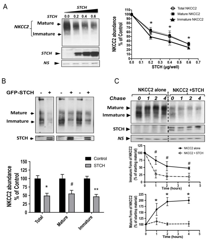
Figure 3.
STCH alters NKCC2 stability and maturation. (A) Total NKCC2 protein abundance is reduced by STCH in a dose-dependent fashion. HEK cells were co-transfected with Myc-NKCC2 (0.2 μg/well) and increasing amounts of STCH (0.2–0.6 μg/well) as indicated. NKCC2 proteins were detected by Western blotting with Myc antibody (left panel). Right panel, densitometric analysis of total, immature, and mature NKCC2 proteins. Data are expressed as a percentage of control. *, p < 0.05 (n = 3). (B) STCH co-expression decreases the expression of NKCC2 proteins. Upper panel, representative immunoblot analysis showing the effect of STCH overexpression on NKCC2 protein abundance in HEK cells. Cells were transfected with Myc-NKCC2 alone (0.2 μg/well) or in the presence of GFP-STCH (0.6 μg/well). 16–18 h post-transfection, total cell lysates were subjected to immunoblot analysis for Myc-NKCC2 and anti-GFP. Lower panel, quantitation of steady state mature, immature, and total NKCC2 expression levels with or without STCH co-expression. Data are expressed as a percentage of control ± SE, *, p < 0.04 (n = 4); #, p < 0.003 (n = 4); **, p < 0.002 (n = 4), versus control. (C) STCH decreases NKCC2 stability and maturation. Upper panel, representative immunoblot showing cycloheximide chase analysis of NKCC2 in the presence or absence of GFP-STCH. 14–16 h post-transfection, HEK cells transiently expressing WT NKCC2 alone or in combination with STCH, were chased for the indicated time after addition of cycloheximide. Total cell lysates were separated by SDS-PAGE and probed by anti-Myc antibodies. Lower panels, quantitative analysis of NKCC2 stability and maturation. The density of the mature and immature form of NKCC2 proteins was normalized to the density at time 0. #, *; p < 0.05 (n = 3) versus control. NS, a non-specific band illustrating the equal loading of protein extracts.

