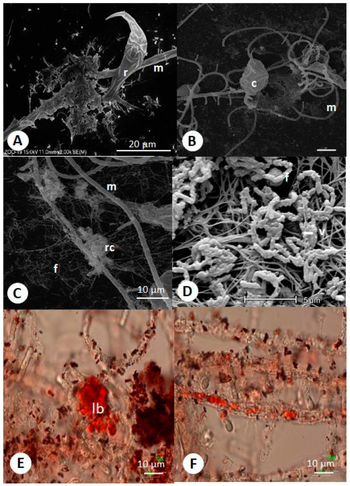
Figure 3.
SEM (scanning electron microscopy) and LM (light microscopy) micrographs of Zoophagus insidians (predatory fungus), rotifers (Lecane inermis), and bacteria cultured in “Żywiec” water. (A) Rotifer attached to glue formed fungal and bacterial material and fungus on the glass surface; (B) rotifer trapped by fungus; (C) remnants of rotifers after digestion by fungi and bacteria; (D) bacteria with virus-like particles; (E) lipid bodies in rotifer stained with oil red O; (F) lipids transported from rotifer to mycelium; r—rotifer; c—immobilized rotifer; rc—remnants of cadaver and absorptive hyphae; m—mycelium; f—flagella-like structures; v—virus-like particles; Ib—lipid bodies.

