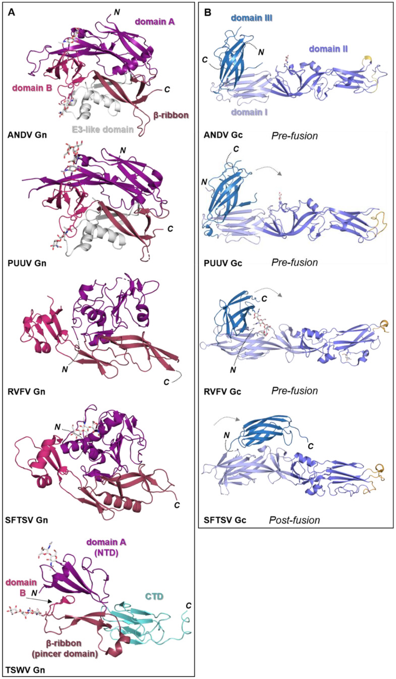
Figure 5.
Known structural features of bunyaviral Gn and Gc envelope proteins. (A) The Gn envelope glycoprotein displays limited structural similarity across bunyavirus families. Five crystal structures of Gn ectodomain regions of different bunyaviruses are shown. Upper panel: cartoon representation of the Gn N-terminal region of the ectodomain of the New World orthohantavirus ANDV (PDB: 6Y6P [154]), which displays a four-domain architecture (domain A, deep purple; a β-ribbon domain, purple-brown; domain B, warm pink; and a domain reminiscent of the alphavirus E3 protein, white). Second panel: cartoon representation of the Gn N-terminal region of the ectodomain of the Old World orthohantavirus PUUV (PDB: 5FXU [165]). Third panel: cartoon representation of the N-terminal region of the Gn ectodomain from RVFV (PDB: 6F8P [151]). Fourth panel: cartoon representation of the N-terminal region of the Gn ectodomain from SFTSV (PDB: 5Y10 [166]). Interestingly, SFTSV Gn contains a region reminiscent of the E3-like domain observed in hantavirus Gn proteins. Bottom panel: cartoon representation of the Gn ectodomain from TSWV (PDB: 6Y9L [167]). TSWV Gn displays a largely conserved three-domain architecture in which domain B is reduced to a β-hairpin. The C-terminal domain (CTD) comprises a β-sandwich domain (cyan) (please see Figure 6). (B) Structurally characterized bunyaviral Gc fusion proteins display a conserved class II fusion protein architecture (domain I, light blue; domain II, slate blue; domain III, sky blue). Four crystal structures of the Gc ectodomain of different bunyaviruses are shown in a putative pre-fusion conformation (except SFTSV Gc for which a post-fusion state was determined). The dashed grey arrow indicates the movement of domain III between putative pre- and post-fusion conformations. Fusion loop(s) are indicated in bright orange. Top panel: crystal structure of the ANDV New World orthohantavirus Gc protein ectodomain in its pre-fusion conformation (PDB: 6Y5F [154]). Second panel: crystal structure of the Old World orthohantavirus PUUV Gc protein ectodomain in its pre-fusion conformation (PDB: 7B09 [168]). Third panel: crystal structure of the RVFV phlebovirus Gc protein ectodomain in its pre-fusion conformation (PDB: 4HJ1 [160]). Bottom panel: crystal structure of the SFTSV Gc protein ectodomain in its post-fusion conformation (PDB: 5G47 [169]). Note that the position of domain III has shifted from the tip of domain I in pre-fusion conformations towards the border of domains I and II in this post-fusion state. In all structural representations crystallographically observed glycans are shown as white sticks.

