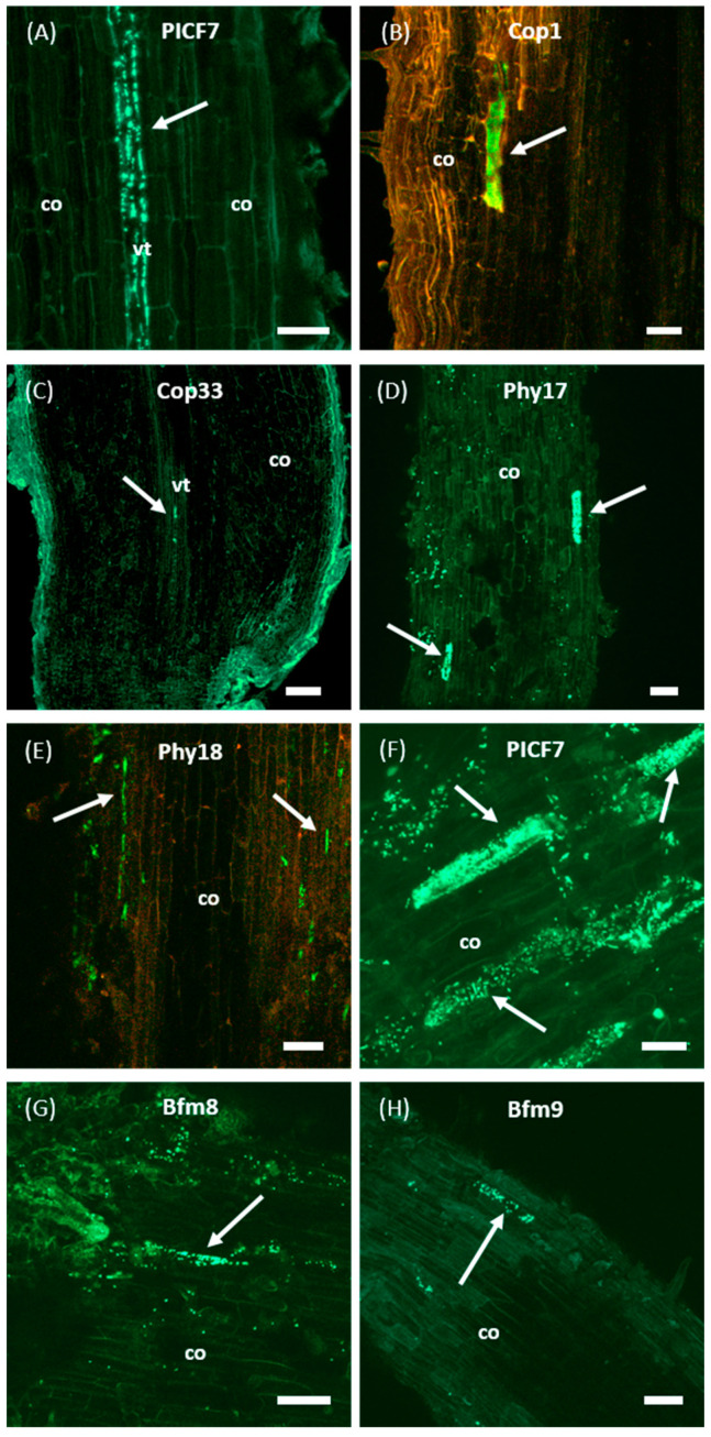
Figure 5.
Confocal laser scanning microscopy images of olive (cultivar Picual) roots colonized by GFP-labeled Pseudomonas simiae PICF7 and its selected Tn5-TcR insertion mutants. Images show inner colonization events of different root tissues by wild-type PICF7 (panel (A)), and mutants Cop1 (B), Cop33 (C), Phy17 (D) and Phy18 (E). Additionally, colonization of olive root surface by wild-type PICF7 (panel (F)) and biofilm mutants Bfm8 (G) and Bfm9 (H) are also shown. Images are representative of the colonization events observed and were taken from 4 to 17 days after root bacterization with fluorescently labelled derivatives. Scale bar represents 50 μm in all panels, except in ((C), 100 μm) and ((F), 20 μm). White arrows point to spots or microcolonies of the inoculated bacteria. co, cortical cells; vt, vascular tissue. In panels (B,E), the red channel was added to increase plant tissue contrast and improve visualization.

