Figure 1.
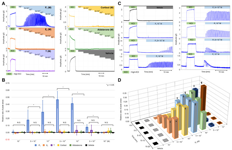
Effects of various natural steroid hormones on the contractile activities in the resting myometrium. (A) Representative myograph of six myographs obtained from experiments that used myometrial strips prepared from the uterine tissues of rats at 20 days of gestation. Progesterone (P4), 17β-estradiol (E2), testosterone (T), cortisol, and aldosterone were added sequentially to a physiological saline solution (PSS) at concentrations of 10−7 M–10−4 M. (B) The relative ratio of the peak area (RRPA) of the progesterone (P4), 17β-estradiol (E2), testosterone (T), cortisol, aldosterone, and vehicle groups in the resting myometrium. The peak area of high-KCl-induced contractions for 10 min was defined as the reference area. (C) Representative myograph of six myographs obtained from experiments that evaluated the effects of single-dose progesterone on the contractile activities in the resting myometrium. Progesterone was added to the PSS at the concentrations of 10−7–10−4 M. (D) Time- and concentration-dependent changes in the RRPA of the vehicle and progesterone groups at various progesterone concentrations. The peak area of high-KCl-induced contractions for 5 min was defined as the reference area. * p < 0.05, † p < 0.05, ‡ p < 0.05, § p < 0.05, ‖ p < 0.05, ¶ p < 0.05.

