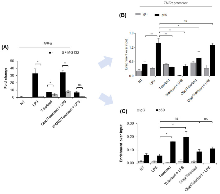
Figure 3.
PARP1 eviction from the TNF-α promoter prevents p65 re-binding after LPS re-stimulation. (A) The contribution of canonical NF-κB to macrophage response to LPS was estimated by comparing TNF-α transcription in control and cells pre-treated with proteasome inhibitor (MG132; 1 µM). mRNA level of the cytokine was quantified by real-time PCR, normalized to median of ACTB and GAPDH and presented as a fold change with respect to untreated cells. (B) p65 and (C) p50 occurrence at the TNF-α promoter was analyzed by ChIP-real-time PCR. Bars in the figures represent mean ± standard error of the mean (SEM). One-way analysis of variance (ANOVA1) was carried out in GraphPad Prism 5 to compare means in several groups. Once the significance was detected, ANOVA1 was followed by the Tukey post-hoc test and significant differences between the two considered means are marked with * when significant at p < 0.05, ** when significant at p < 0.01, ns—non-significant at p > 0.05. Abbreviations: LPS—lipopolysaccharide, Olap—olaparib, TNF—tumor necrosis factor, ACTB—actin beta, GAPDH—glyceraldehyde 3-phosphate dehydrogenase, IgG—immunoglobulin G, p65—transcription factor p65 (RelA), p50—nuclear factor NF-kappa-B p105 subunit.

