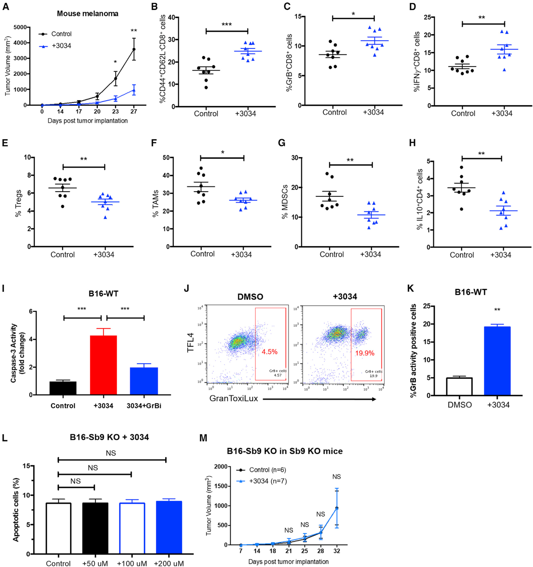
Figure 5. Treatment with a Small Molecule Inhibitor of Sb9 Suppresses Melanoma Progression In Vivo.

(A) Tumor growth curve demonstrates significantly slower growth of B16-WT tumors in the WT mice treated with Compound 3034 (300 μg intraperitoneally [i.p.] bid for 14 days following implantation) (n = 17) than the control group treated with 10% DMSO (n = 17). *p < 0.05, **p < 0.01.
(B–D) Flow cytometric analyses demonstrate significantly higher percentages of tumor-infiltrating CD44highCD62LlowCD8+ effector memory T cells (B), GrB+CD8+T cells (C), and IFNγ +CD8+T cells (D) in tumors from the 3034-treatment group than the control at day 17 post-implantation. *p < 0.05, **p < 0.01, ***p < 0.001.
(E–G) Flow cytometric analyses show significantly lower percentages of tumor-infiltrating CD4+CD25+Foxp3+ Treg populations (E), CD45+CD11b+F4/80+Ly6C−Ly6G− TAM populations (F), and CD45+CD11b+Gr1+CD3− MDSC populations (G) in tumors from the 3034-treatment group than the control at day 17 post-implantation. *p < 0.05, **p < 0.01.
(H) Flow cytometric analysis shows significantly lower percentages of tumor-infiltrating IL-10+CD4+population in tumors from the 3034-treatment group than the control at day 17 post-implantation. **p < 0.01.
(I) Caspase-3 activity assay demonstrates that compound 3034 (200 μM) induces GrB-mediated apoptosis in B16 cells, as determined by the EnzChek Caspase-3 Assay Kit. The GrB inhibitor (368050) was used at 10 μM. ***p < 0.001.
(J and K) GranToxiLux assay demonstrates increased activity of GrB in B16 cells treated with compound 3034 (200 μM) for 24 h. **p < 0.01.
(L) Quantitative flow cytometric analysis shows that compound 3034 does not induce additional apoptosis at various concentrations (50 μM, 100 μM, and 200 μM) in the B16-Sb9 KO cells, as indicated by annexin-V+ and viability dye− population. NS, no significant difference.
(M) Tumor growth curve indicates no difference in antitumor effect of compound 3034 on B16-Sb9 KO tumors in Sb9 KO mice (n = 7), as compared to the control (n = 6). NS, no significant difference. See also Figure S5.
