FIGURE 1:
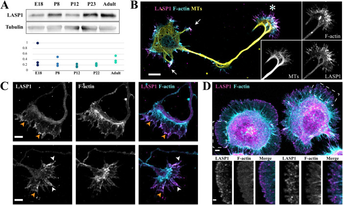
LASP1 is expressed in the developing brain and localizes to the axon growth cone. (A) Top, a representative Western blot showing the developmental profile of LASP1 in rat hippocampi at the indicated stages. Bottom, the plot shows normalized LASP1 levels relative to tubulin loading control from three independent blots. (B) Representative confocal images of a rat hippocampal neuron in culture at 2 d in vitro (DIV2) stained for endogenous LASP1 (magenta), F-actin (phalloidin, cyan), and α-tubulin (yellow). Individual channels of the growth cone are shown as the insets on the right. Arrows indicate the minor processes destined to be dendrites, and asterisk indicates the axonal growth cone. Scale bar: 20 µm. (C) Representative confocal images of growth cones from fixed DIV2 cultured rat hippocampal neurons, stained for endogenous LASP1 (magenta) and F-actin (phalloidin, cyan). LASP1 localizes to the leading edge of lamellipodia (orange arrowheads) and actin bundles in filopodia (white arrowheads). Scale bars: 5 µm. (D) Representative confocal images of two CAD cells labeled for LASP1 (magenta) and F-actin (phalloidin, cyan). LASP1 is seen at lamellipodia and filopodia in a similar pattern to growth cones. Scale bars: 5 µm (main panel) and 2 µm (insets).

