FIGURE 6:
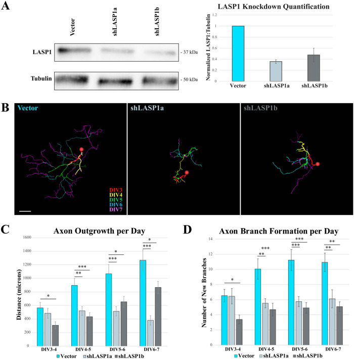
Knockdown of LASP1 truncates axon elongation and reduces branching in cultured hippocampal neurons. (A) Representative anti-LASP1 and anti-tubulin Western blots of lysates from hippocampal neurons transfected with empty plasmid (vector), shLASP1a, or shLASP1b for 72 h. Graph shows ∼60–70% reduction in LASP1 expression in cultures expressing shRNAs across three independent culture replicates. LASP1 levels are normalized to tubulin loading controls. Error bars represent standard error. (B) Axon tracings of representative rat hippocampal neurons transfected at DIV2 with empty backbone control (Vector), shLASP1a, or shLASP1b. Neurons were imaged every 24 h from DIV3 until DIV7, and each day’s growth is temporally color coded (scale lower right, Vector image). Knockdown of LASP1 reduces axon outgrowth over each 24 h period. Scale bar: 150 µm. (C) Graph depicting axon outgrowth (in µm) every 24 h. (D) Graph depicting the number of newly formed branches at each time point. Error bars represent standard error. *p < 0.05; **p < 0.005; ***p < 0.001. P values calculated using a one-way ANOVA Tukey’s HSD post-hoc test. Data collected from approximately 30 neurons from three independent cultures (see Supplemental Tables S1 and S2 for exact n and p values).

