FIGURE 8:
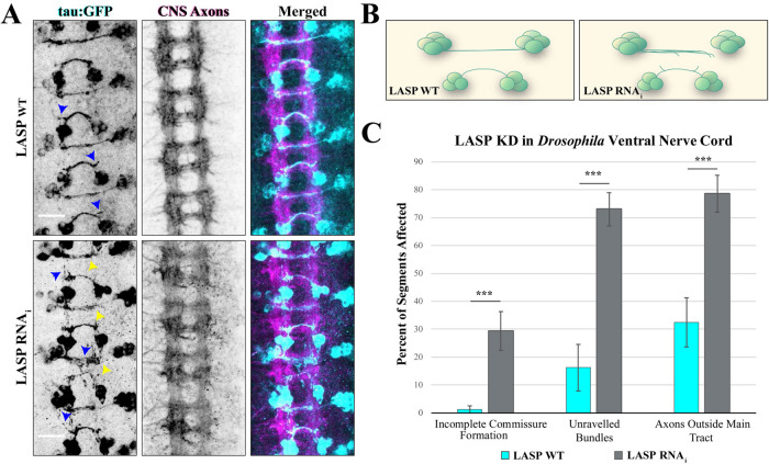
Lasp knockdown leads to defects in axon commissure formation in Drosophila. (A) Representative images of stage 16 Drosophila embryos labeled with tau:GFP in egl-expressing ventral nerve cord axons. Top row shows dicer-only control embryo (WT-LASP); bottom row shows embryo expressing dicer plus LASP RNAi (LASP KD). Left column shows GFP-expressing egl neurons (green), middle column shows CNS axon (BP102, magenta), right column shows merged images. Arrowheads indicate defects in commissural axon development, including defasciculation (blue) and axon tracts that do not reach their targets (yellow). Scale bars: 15 µm. (B) Schematic of one segment from wild-type (left) or LASP knockdown (right) egl neurons in the Drosophila ventral nerve cord. Lasp knockdown results in numerous guidance defects, including defasciculation of axon bundles and truncated commissures. (C) Graph shows axon guidance defects from 10 control and 18 knockdown embryos collected across three independent experiments. ***p < 0.001; both defasciculation measures were analyzed using a Student’s t test. Incomplete commissure formation calculated using a Welch’s t test. Error bars represent standard error.

