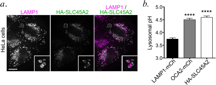
FIGURE 3:
HA-SLC45A2 expressed in HeLa cells localizes to lysosomes and neutralizes lysosomal pH. (a). HeLa cells transiently transfected with HA-SLC45A2 were fixed, labeled for HA (green) and the lysosomal protein LAMP1 (magenta), and analyzed by dIFM. Merged image is at right. Insets of boxes are magnified fivefold. Scale bar, 10 μm. (b) HeLa cells transiently transfected with LAMP1-mCherry alone, mCherry-OCA2, or HA-SLC45A2 and LAMP1-mCherry were incubated with LysoSensor DND-160 and analyzed by fluorescence microscopy with excitation wavelength 405. The W1/W2 emission ratio for mCherry-labeled endolysosomes was calculated and compared with that from a standard curve to define pH as described in Materials and Methods. Data represent mean ± SEM from three independent experiments with the following total sample sizes: 319 LAMP1-mCherry+ endolysosomes, 391 OCA2-mCherry+ endolysosomes, and 299 HA-SLC45A2/LAMP1-mCherry+ endolysosomes. Statistical significance was determined using Welch’s ANOVA with Games-Howell test for multiple comparisons; only significant differences are indicated. ****p < 0.0001.

