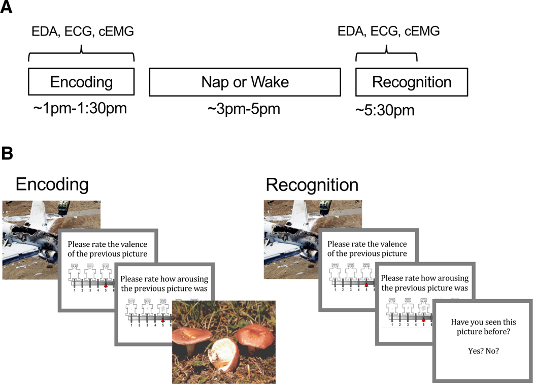
Fig. 1.
Experimental procedure and task. (A) Encoding took place in the early afternoon followed by a 2-hr nap opportunity or wake period and then Recognition. Electrodermal activity (EDA), electrocardiography (ECG), and corrugator supercilii electromyography (cEMG) were measured during the entirety of Encoding and the first half of Recognition. (B) During Encoding participants viewed 90 pictures (targets) and rated the valence and arousal of each on 9-point self-assessment manikin scales. During Recognition, participants viewed 180 pictures, a mixture of target and novel foil pictures, and rated each one on valence and arousal. Participants indicated whether or not they recognized the picture by responding yes/no.

