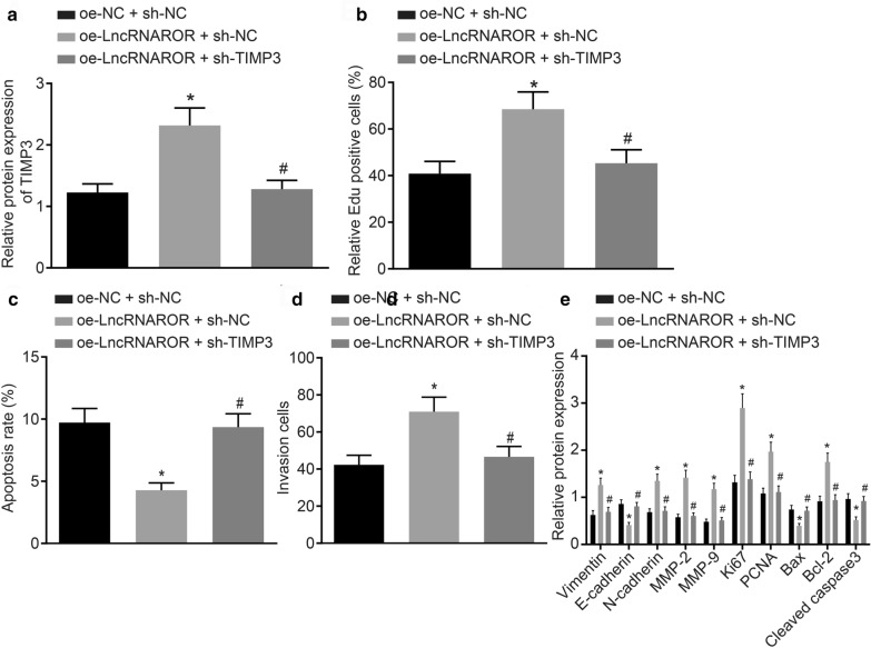
Fig. 4.
The effects of lncRNA ROR on the proliferation, invasion and apoptosis of breast cancer cells are reversed by TIMP3 silencing. a The expression of TIMP3 in MCF-7 cells co-transfected with oe-lncRNA ROR and/or sh-TIMP3 determined by Western blot assay. b The proliferation of MCF-7 cells co-transfected with oe-lncRNA ROR and/or sh-TIMP3 detected by EdU assay. c The apoptosis of MCF-7 cells co-transfected with oe-lncRNA ROR and/or sh-TIMP3 detected by flow cytometry assay. d The invasion of MCF-7 cells co-transfected with oe-lncRNA ROR and/or sh-TIMP3 detected by Transwell assay and the expression of TIMP3 of MCF-7 cells transfected with oe-lncRNA ROR and/or sh-TIMP3 determined by Western blot analysis. e The protein levels of Vimentin, N-cadherin, MMP-2, MMP-9, Ki67, PCNA, Bcl-2, E-cadherin, Bax and Cleaved caspase-3 in MCF-7 cells transfected with oe-lncRNA ROR and/or sh-TIMP3 determined by Western blot analysis. *p < 0.05 vs. that of the cells co-transfected with oe-NC and sh-NC. #p < 0.05 vs. that of the cells co-transfected with oe-lncRNA ROR and sh-NC. The above data are measurement data and expressed as mean ± standard deviation. Comparisons among multiple groups are analyzed by independent sample t test. The experiment is repeated 3 times. MLL1, mixed-lineage leukemia 1; TIMP3, tissue inhibitors of metalloproteinase 3; NC, negative control; FISH, fluorescent in situ hybridization; DMSO, dimethyl sulfoxide; RIP, RNA immunoprecipitation; ChIP, Chromatin immunoprecipitation

