
FIGURE 4.
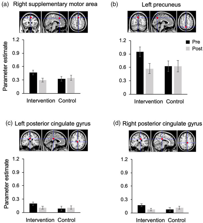
The parameter estimates obtained from fMRI analyses for the VWM task in the (a) right supplementary motor area, (b) left precuneus, (c) left posterior cingulate gyrus and (d) right posterior cingulate gyrus. These four regions showed significant group‐by‐time interactions. VWM, visual working memory