
Figure 1.
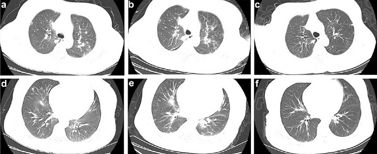
Representative chest computed tomography (CT) scans of this patient. a, d) Chest CT scan obtained at admission showed multiple ground-glass opacities with uneven density involving both upper lungs and right lung. b, e) Chest CT obtained on February 25 before CP transfusion, where multiple shadows of high density in both upper lungs and patchy consolidation in the right lung were observed. c, f) CT image taken on March 6 showed the absorption of bilateral ground-glass opacity after CP transfusion.