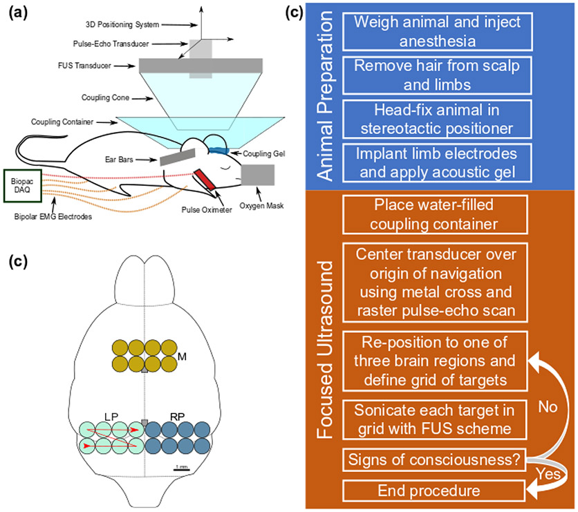
Fig. 1.
Experimental setup. (a) The focused ultrasound system and signal acquisition equipment are labelled on the diagram of a head-fixed mouse in a stereotactic positioner. (b) An outline of the mouse brain is shown with the three brain regions investigated (M: motor, LP: left posterior, RP: right posterior). The red line denotes the sequence of target sonication from 1 to 8. Two grey boxes along the midline denote the bregma and lambda landmarks from top to bottom, respectively. (c) A general outline of the experimental procedure.

