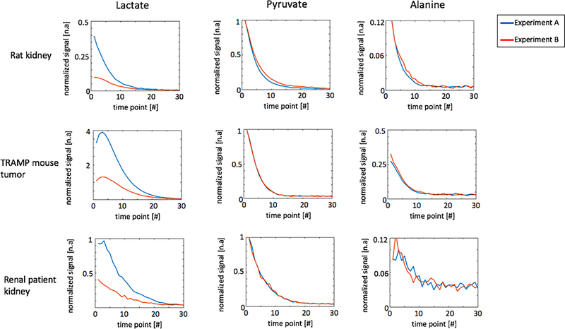
Figure 7:
Representative dynamic curves of lactate, pyruvate and alanine signals acquired in experiment A (pyruvate and alanine: MS-GRE; lactate: MS-3DSSFP) and experiment B (pyruvate, lactate and alanine: MS-GRE). Experiment parameters are described in Table 1. All signals were divided by corresponding noise signals and then divided by the highest value of the pyruvate dynamic curve. Corresponding dynamic images are shown in Supporting Information Figure S4, Supporting Information Figure S5 and Supporting Information Figure S6.

