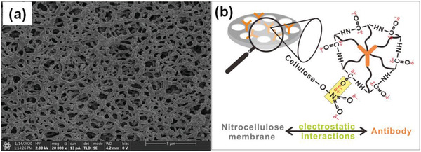
Figure 3.

a) Scanning electron microscopy (SEM) image of nitrocellulose membrane, which indicates the homogeneous porous structure with high surface area. b) Schematic illustration for the interaction mechanism between the carbonyl groups of antibody and the nitrate groups of nitrocellulose membrane.
