
Figure 4.
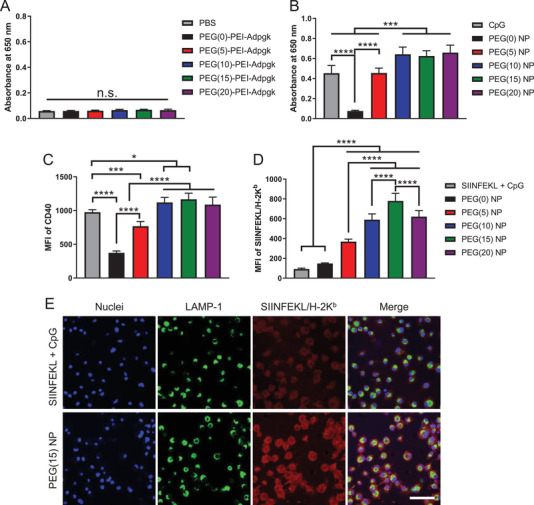
Induction of TLR9‐mediated immune stimulation and antigen cross‐presentation by nanovaccines. A,B) HEK‐Blue TLR9 cells were incubated with A) free polymer form of PEG–PEI–Adpgk conjugates or B) their nanovaccines with CpG, and induction of TLR9 signaling cascade was quantified using 650 nm absorbance. Upregulation of C) CD40 and D) SIINFEKL/H‐2Kb expression by BMDCs after 24 h incubation with SIINFEKL + CpG or SIINFEKL nanovaccines. E) Confocal microscope images of BMDCs incubated with SIINFEKL + CpG or PEG(15) NP of SIINFEKL nanovaccine. Scale bar = 50 µm. The data show mean ± s.d. (n = 6). *P < 0.05, ***P < 0.001, and ****P < 0.0001, analyzed by one‐way ANOVA with Bonferroni multiple comparisons post‐test.