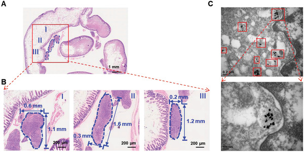
Figure 6.

The H&E staining and TEM images of the tissue from post‐surgery site of orthotopic SKOV3 tumor model. A) The low magnification microscope photo of the tissue from the surgery site. B) The high magnification microscope photo of tumor regions from (A), the blue dash line outlined the residue micro‐tumors of the tissue post‐surgery. C) TEM images of the tissues, which showed presence of nanoparticles (red boxes) in post‐operative tumor residues.
