Figure 5.

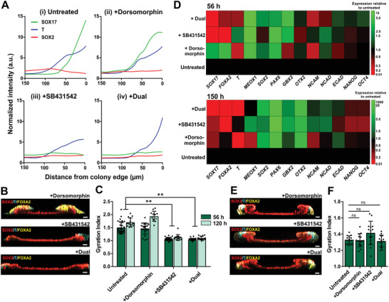
Specification of endodermal cells by TGF signaling supported prospective NE tissue folding. A) Spatial expression profiles of SOX17, T, and SOX2 in micropatterned H9 derived hPSC colonies at 34 h after differentiation in (i) untreated controls or in the presence of (ii) 2 µm Dorsormorphin; (iii) 1 µm SB431542; (iv) 2 µm Dorsormorphin + 1 µm SB431542. All inhibitors were added after 10 h of ME induction. B,C) Inhibition of TGFβ signaling initiated at 10 h post induction resulted in the specific abrogation of endodermal (FOXA2+) lineages, which was accompanied by attenuation of tissue folding. B) Cross‐sectional view of 3D confocal sections of whole 56‐h old ME‐primed μNETs treated with inhibitors. C) Gyration indices of 56‐h and 120‐h old ME‐primed μNETs. Data are average ± s.e.m of at least eight colonies from three independent experiments. D) Heat map displaying transcriptional profiles of 56‐h old and 150‐h old ME‐primed μNETs treated with different inhibitors initiated at 10 h after induction relative to untreated control samples. Transcript levels are average of three independent experiments. E,F) Addition of inhibitors at 34 h after induction when patterning of three germ lineages have been established did not affect tissue folding. E) Cross‐sectional views of 3D confocal sections of whole 56‐h old ME‐primed μNETs treated with inhibitors, and corresponding F) gyration indices indicative of tissue folding. Data in (F) are average ± s.e.m of 7 colonies from two independent experiments. Asterisks indicate statistical significance (One‐way ANOVA followed by Tukey´s post‐test, **p < 0.0001; n.s. indicates no statistical significance). Scale bars in (B) and (E) = 100 µm.
