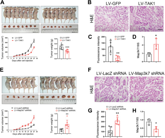
Figure 3.

TAK1 negatively regulates esophageal squamous tumor cell growth in vivo. A) ECA‐109 cells were transduced with lentivirus expressing TAK1 (LV‐TAK1) or GFP (LV‐GFP). After selection using puromycin, the cells were implanted into nude mice. Tumor volume and weight were reduced with the LV‐TAK1 transduced cells. n = 8. B) H&E staining of the tumors transduced with LV‐TAK1. Scale bar = 200 µm. C) Quantitative analysis of immunostaining of Ki67 in the tumors transduced with LV‐TAK1. n = 5. D) qRT‐PCR analysis of TAK1 expression in LV‐TAK1 transduced tumors. n = 3. E) ECA‐109 cells were transduced with lentivirus bearing Map3k7 shRNA (LV‐Map3k7 shRNA) or LacZ shRNA (LV‐LacZ shRNA). After selection using puromycin, the cells were implanted into nude mice. Tumor volume and weight were improved with the LV‐Map3k7 shRNA transduced cells. n = 6, 7. F) H&E staining of the tumors transduced with LV‐Map3k7 shRNA. Scale bar = 200 µm. G) Quantitative analysis of Ki67 staining of the tumors transduced with LV‐Map3k7 shRNA. n = 5. H) qRT‐PCR analysis of TAK1 expression in tumors transduced with LV‐Map3k7 shRNA. n = 3. Data are presented as mean ± SD (error bars). Statistical significance was tested by two‐tailed unpaired Student t‐test. *p < 0.05, **p < 0.01, and ***p < 0.001.
