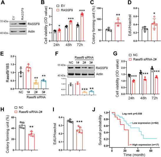
Figure 5.

RASSF9 positively regulates esophageal squamous tumor cell proliferation. A) ECA‐109 cells were transfected with plasmid expressing RASSF9. Western blot analysis showing RASSF9 was increased following the transfection. Actin was used as a loading control. Representative blots were shown (n = 3 biologically independent replicates per group). B–D) Elevated expression of RASSF9 stimulates cell viability (B; n = 9 biologically independent replicates per group), colony formation (C; n = 4 biologically independent replicates per group), and EdU incorporation (D; n = 5 biologically independent replicates per group) in ECA‐109 cells. E) ECA‐109 cells were transfected with siRNAs targeting Rassf9. Rassf9 siRNAs decreased mRNA levels of Rassf9 (n = 3 biologically independent replicates per group). Gene expression was analyzed by qRT‐PCR and 18S was used for normalization of the gene expression. F) Protein level of RASSF9 was reduced by Rassf9 siRNAs in ECA‐109 cells (n = 3 biologically independent replicates per group). Protein level was analyzed by western blotting and Actin was used a loading control. Representative blots were shown (n = 3 biologically independent replicates per group). G–I) Knockdown of RASSF9 decreases cell viability (G; n = 9 biologically independent replicates per group), colony formation (H; n = 4 biologically independent replicates per group), and EdU incorporation (I; n = 5 biologically independent replicates per group) in ECA‐109 cells. J) TCGA database showing that RASSF9 expression negatively correlates with survival time in ESCC patients. Data are presented as mean ± SD (error bars). Statistical significance was tested by two‐tailed unpaired Student t‐test (B–I) or log‐rank (Kaplan–Meier) test (J). *p < 0.05, **p < 0.01, and ***p < 0.001.
