Figure 7.

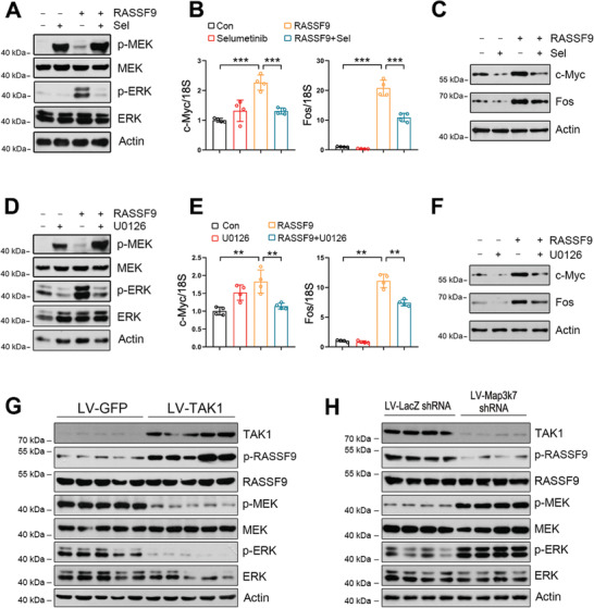
MEK inhibition counteracts RASSF9‐induced ERK activation and oncogene expression. A) MEK inhibition by selumetinib reduces RASSF9‐induced ERK activation. B–C) MEK inhibition by selumetinib decreases the expression of c‐Myc and Fos at B) mRNA and C) protein levels. ECA‐109 cells were transfected with plasmid carrying RASSF9. The cells were treated with 10 µm of selumetinib 12 h post transfection, for additional 24 h. D) MEK inhibition using U0126 abolishes RASSF9‐induced ERK activation. E,F) U0126 blocks RASSF9‐induced the expression of c‐Myc and Fos at E) mRNA and F) protein levels. Cell treatments were similar to the procedures in (A–C) except that 10 µm U0126 was used instead of 10 µm selumetinib. G,H) Protein levels of TAK1, p‐RASSF9, RASSF9, p‐MEK, MEK, p‐ERK, ERK in the transplanted tumors in nude mice. The transplantation procedures were described in Figure 3. n = 5. Protein and mRNA levels were analyzed by western blot analysis and qRT‐PCR, respectively. Actin was used as a loading control. 18S was used for normalization of the gene expression in qRT‐PCR. Sel: selumetinib. Sample size for data in (A, C, D, F) is n = 3 biologically independent replicates per group and representative blots were shown. Sample size for data in (B, E) is n = 4 biologically independent replicates per group. Data are presented as mean ± SD (error bars). Statistical significance was tested by two‐tailed one‐way ANOVA test. **p < 0.01 and ***p < 0.001.
