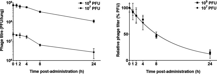
FIG 3.
Phage titer in the lungs (lung tissues and BALF combined) of healthy mice after intratracheal administration of phage PEV31 at doses of 107 and 109 PFU. Phage titer is expressed as PFU per lung homogenate and BALF combined (left) and as PFU relative to the administered dose (right). Lines of regression (superimposed) are shown in the right panel. Error bars denote standard deviation (n ≥ 4 except for t = 2 h of the 107 PFU group and t = 1 h and 4 h of the 109 PFU group, for which n = 3).

