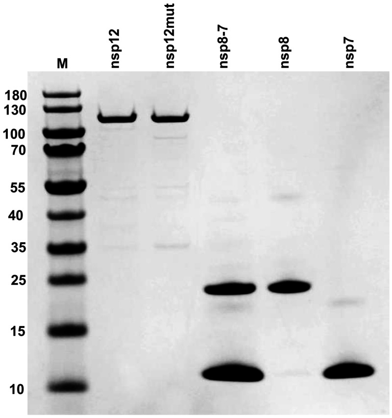
FIG 1.
nsp12, nsp7, nsp8, and nsp8-7 PAGE protein gel stained with Coomassie blue. nsp12mut contains an active-site mutation (active-site motif SDD changed to SAA). nsp8-7 represents copurified nsp8 and nsp7. M, protein molecular weight markers. The sizes of protein markers (in kilodaltons) are indicated on the left.

