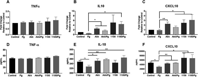
FIG 4.
A. muciniphila and Amuc_1100 increase the levels of anti-inflammatory mediators in the gingival tissue during P. gingivalis infection. (A to C) Gingival tissues isolated at the endpoint of the gavage period were analyzed for mRNA expression. Mice were divided into the following groups: PBS (control), P. gingivalis (Pg), A. muciniphila (Akk), P. gingivalis and A. muciniphila (Akk/Pg), Amuc_1100 (1100), and Amuc_1100 and P. gingivalis (1100/Pg). The relative expression of TNF-α (A), IL-10 (B), and CXCL10 (C) mRNAs was determined by quantitative real-time PCR using TaqMan assays. The fold change was calculated using control tissue as a baseline and β-actin as a normalizing control. (D to F) Gingival tissue lysates were also analyzed for the protein concentrations of TNF-α (D), IL-10 (E), and CXCL10 (F) via ELISA. All values are represented as means ± SEM (n, 6/group). Results were analyzed by one-way ANOVA, with differences considered significant at a P value of ≤0.05. *, P ≤ 0.05; **, P ≤ 0.01; ***, P ≤ 0.001. (Asterisks indicate comparisons with P. gingivalis; tildes indicate comparisons with the control.)

