Figure 2.
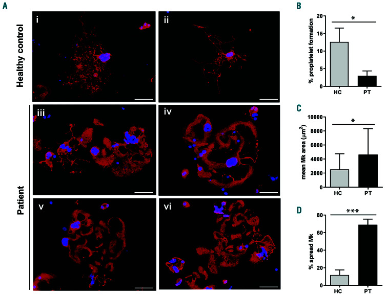
Analysis of proplatelet formation and spreading in adhesion to fibrinogen of megakaryocytes of the investigated patient. Megakaryocytes (Mk) were incubated on fibrinogen-coated coverslips for 16 hours at 37°C and 5% CO2, fixed and stained with an anti-β1-tubulin antibody (red). Hoechst (blue) was used for counterstaining nuclei. (A-D) Mk of the patient exhibited a markedly increased spreading, of ten with aberrant morphology, associated with defective extension of typical proplatelets. (A) (iii-vi) Representative examples of patient’s Mk. (i-ii) Proplatelets-forming Mk of healthy controls processed in parallel are shown for comparison. Scale bars: 60 mm. (B) Proplatelet formation was quantif ied using fluorescence microscopy as the proportion of Mk displaying at least one proplatelet with respect to the total number of Mk. (C,D) Spreading was measured through image analysis as the average area covered by each Mk (C), and as the percentage of spread Mk with respect to the total number of Mk (D). The data repor ted with the histograms are expressed as means ± standard deviation. ***P<0.001, and *P<0.05 by two-tailed Student t-test.

