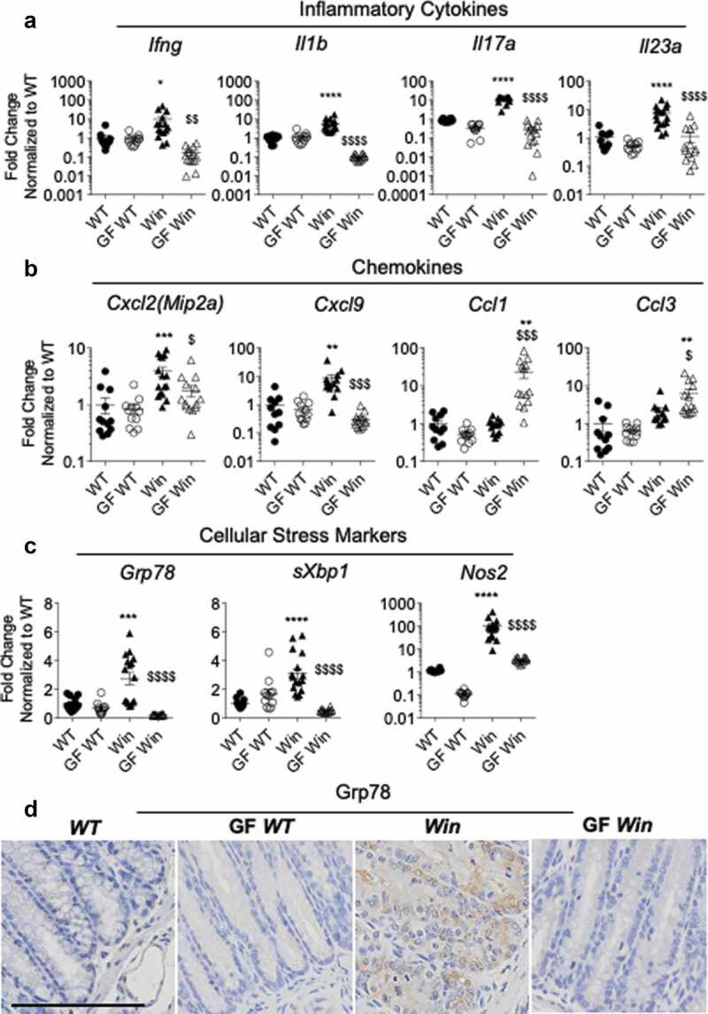
Figure 2.

Inflammation and ER stress were reduced in the distal colon of GF Winnie. (a-c): Distal colonic mRNA expression of genes encoding inflammatory cytokines (a), intestinal epithelial-specific chemokines (b), and cellular stress markers (c) in mice described in Figure 1. (d), Representative immunohistochemistry with Grp78 antibody reflective of ER stress in distal colon sections. (a-c): Data presented as fold change corrected to b actin and normalized to WT mice. Values are expressed as mean ± SEM and individual data points, n = 10 to 14. One-way ANOVA with Bonferroni’s multiple comparison test; *compared to WT control, #compared to GF WT, and $compared to Win (*P < .05; **P < .01; ***P < .001; ****P < .0001)
