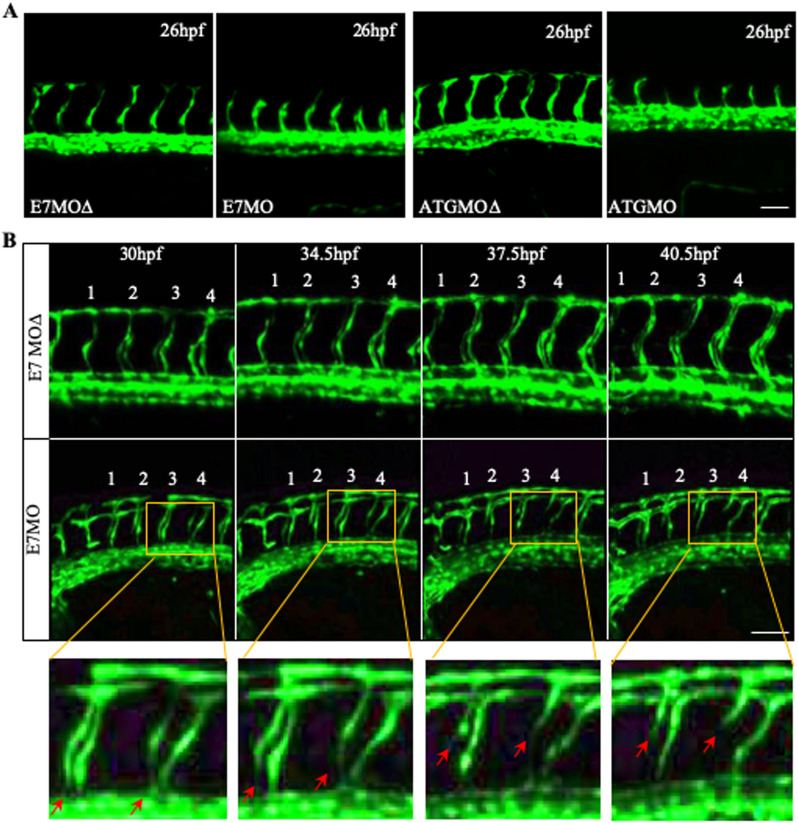
Fig. 3.
Vessel regression in Tspan18 zebrafish morphants. (A) Intact ISV sprouting is observed at 26 hpf in Tspan18-deficient fish. (B) Confocal time-lapse microscopy of regressing ISVs (red arrows) in Tspan18-deficient fish. Numbers 1–4 indicate the order of ISVs. Yellow rectangle: corresponding section shown at higher magnification under the bottom. All larvae lateral view. Anterior is to the left. Scale bars: 50 μm. All experiments (A–B) were repeated at least three times and similar results were obtained.

