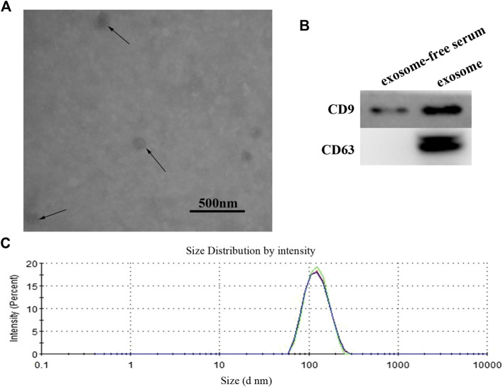
FIGURE 1.
(A) Identification and characterization of exosome. The purified exosome from serum of GC patients was observed under a transmission electron microscope (scale 500 nm). (B) Western blot analysis of CD9 and CD63 expression in free-exosome serum and exosomes isolated from GC serum. (C) The size of exosomes was determined by using the DLS analysis.

