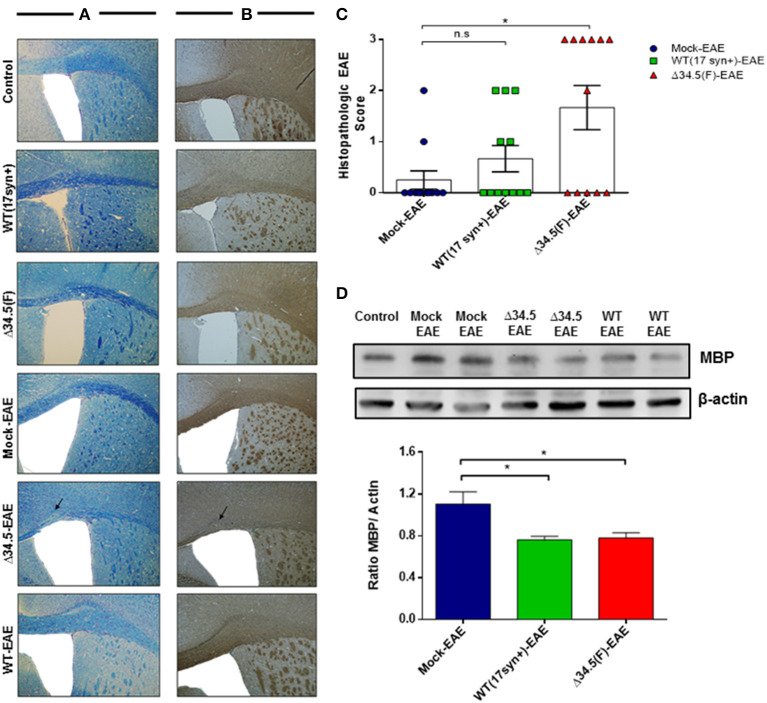
Figure 3.
Asymptomatic HSV-1 infection contributes to brain demyelination after EAE induction. Mice were treated with mock, infected with WT HSV-1 (17syn+ strain) or inoculated with HSV-1 Δ34.5 (F strain) (n = 12/group). EAE was induced 30–35 days post-treatment and 4 mice from each group were euthanized at day 14, 21 or 25 post-EAE induction. (A) Representative images of brain sections stained with Luxol Fast Blue showing the corpus callosum. (B) Representative images of immunohistochemistry against the MBP protein in brain samples. The image magnification is 10× and corresponds to day 14 post-EAE induction. Arrows show demyelination sectors with reduced myelin in the corpus callosum. (C) Quantitative histopathological analyses of brain tissue samples. Values represent means ± SEM of three independent experiments (4 mice/group per day evaluated). Data were analyzed using one-way ANOVA with Dunnett's multiple comparison post-test; **p < 0.05 and n.s. non-significant. (D) Representative western blot images for MBP (upper panel) and β-actin (lower panel) in brain tissue at day 14 post-EAE induction. The graph shows densitometric analyses for MBP bands that were normalized to β-actin. Data represent the mean ± SEM of two independent experiments (n = 6). Comparisons between ratios were performed using one-way ANOVA with Dunnett's multiple comparison post-test; *p < 0.05.

