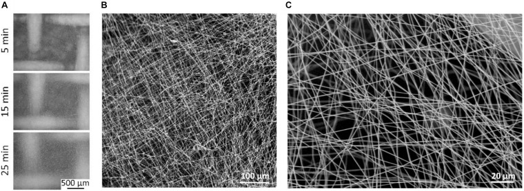
FIGURE 4.
Electrospun substrates by using 15% CA. (A) Optical microscope images of the scaffolds prepared on stainless-steel mesh by using three different spinning times (5, 15, and 25 min). (B) Low magnification and (C) high magnification scanning electron microscopy (SEM) imaging of 25 min electrospun CA scaffolds, as selected candidates for in vitro testing.

