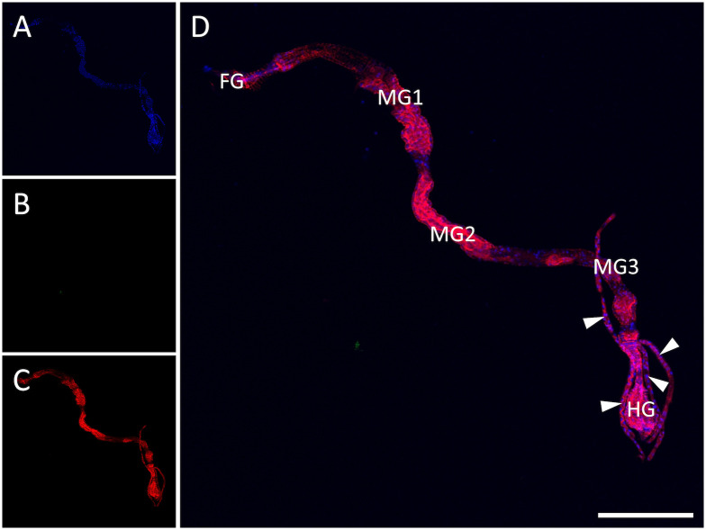
Fig 1. Indirect immunofluorescence detection of watermelon silver mottle virus (WSMoV) in non-viruliferous Thrips palmi.
(A) Nuclei of cells stained with the DNA selective dye DAPI. (B) No WSMoV antigen detected. (C) Actin stained with phalloidin conjugated with Alexa Fluor 633. (D) Merged image, indicating that no WSMoV antigen was detected in the alimentary tract. WSMoV antigen was detected using rabbit antiserum against WSMoV nucleocapsid protein and a secondary antibody conjugated with Alexa Fluor 555. Actin was stained to delineate cell boundaries and tissue types. The confocal image depicting alimentary tract of a thrips is a representative image from 12 non-viruliferous thrips examined in this study. FG: foregut, MG: midgut, HG: hindgut. Arrowheads point to Malpighian tubules. Scale bar = 250 μm.

