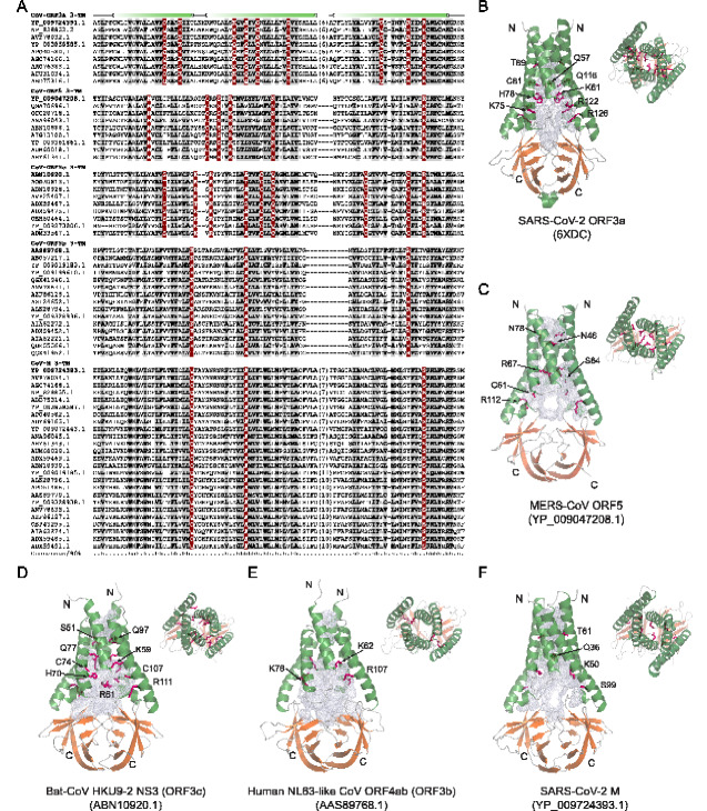
Figure 3.
(A) MSA of 3-TM regions from five CoV ORF3-like (M2) families. The secondary structure is shown above the alignment and the consensus is shown below the alignment, where h stands for hydrophobic residues, s for small residues, b for big residues, and p for polar residues. The numbers are indicative of the excluded residues from sequences. (B–F) Dimeric structures of SARS-CoV-2 ORF3a and homology models of SARS-CoV-2 M, human-NL63-like-CoV ORF3b, bat-CoV HKU9-2 ORF3c, and MERS-CoV ORF5. Both the side-view and top-view are presented. Alpha-helices are colored in green, β-sheets of the β-sandwich domains in orange, and loops in grey. The channel cavities are colored in grey. The polar residues, which are conserved in each family and located on the cavity surface, are shown in a purple stick model. The PDB and NCBI ids are shown in brackets.

