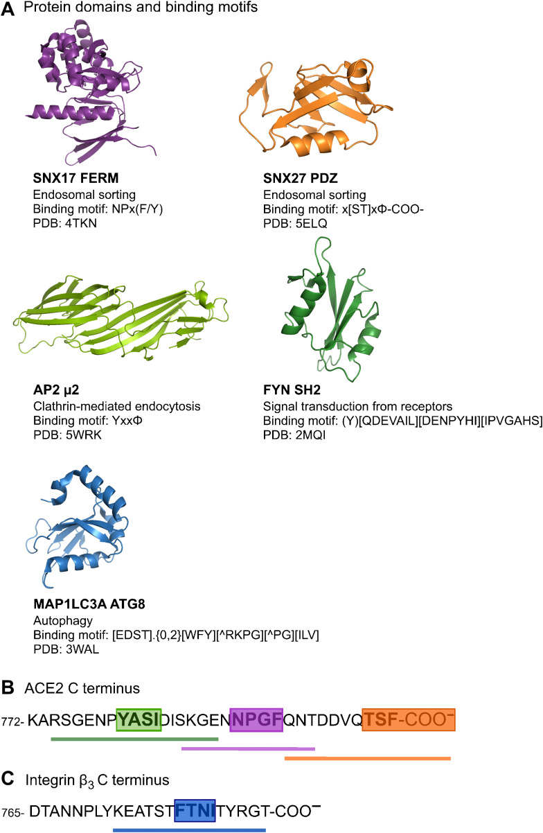
Fig. 1. Overview of domains and peptides tested for binding.
(A to C) Representative structures of peptide-binding domain assessed for their potential interactions with peptide sequences from the cytoplasmic tails of ACE2 (B) and integrin β3 (C). Green: Region containing predicted overlapping binding sites for NCK SH2 domain, the ATG8 domains of MAP1LC3As and GABARAPs, and AP2 μ2. Magenta: Predicted PTB binding site. Orange: Predicted class I PDZ-binding site. Blue: Predicted ATG8 binding site in integrin β3. PDB, Protein Data Bank.

