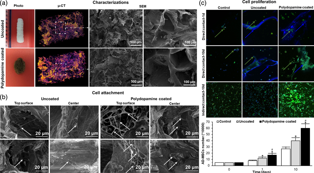
FIGURE 4.
(a) Photographs, μ-CT, SEM images of an uncoated- and polydopamine coated-cryogel bioscaffold: In μ-CT images, the purple areas show the bioscaffold material and the dark areas refer to the void space, SEM images showing the existence of both small, large, and continuously interconnected macropores throughout the entire bioscaffold construct; (b) SEM images of AD-MSCs (indicated by white arrows) on the superficial layer and within the center of uncoated- and polydopamine coated-cryogel bioscaffolds at Day 10; (c) confocal images of AD-MSCs after 1 and 10 days seeding into uncoated- and polydopamine coated-cryogel bioscaffolds (direct contact) and culturing with the medium of uncoated- and polydopamine coated-cryogel bioscaffolds (indirect contact) and the results of AD-MSCs counting at Day 0, 1, and 10 showing the improved AD-MSCs proliferation when cryogel bioscaffolds were coated with polydopamine (reproduced with permission from Razavi, M., Hu, S., & Thakor, A. S. (2018). A collagen based cryogel bioscaffold coated with nanostructured polydopamine as a platform for mesenchymal stem cell therapy. Journal of Biomedical Materials Research Part A, 106, 2213–2228). Significant differences: *p < .05, difference between the control group and bioscaffolds. #p < .05, difference between uncoated- and polydopamine coated-cryogel bioscaffolds. (Unpaired Student’s t-test)

