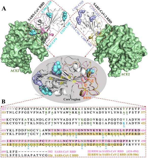
Figure 1.

Crystal structures of proteins acquired from the RCSB PDB and sequence alignment. (A) Structures of SARS-CoV and SARS-CoV-2 RBD–ACE2 complexes. The RBDs are shown in cartoon modes, whereas ACE2 is shown in surface style. The disulfide bonds and RBM are highlighted in cyan and pink in SARS-CoV RBD and blue and yellow in SARS-CoV-2 RBD, respectively. (B) Sequence alignment of SARS-CoV and SARS-CoV-2 RBDs. ‘·’ and ‘*’ represent mutant and key interactional residues, respectively.
