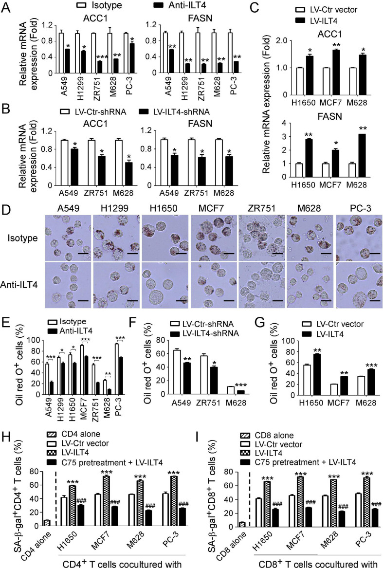
Figure 4.
Tumor-derived immunoglobulin-like transcript 4 (ILT4) promotes T cell senescence via upregulation of tumor lipid metabolism. (A) Neutralization of ILT4 significantly reduced gene expression of key enzymes for fatty acid synthesis (ACC1 and FASN) in tumor cells. Human tumor cell lines were treated with anti-ILT4 neutralizing antibody (500 ng/mL) or isotype control antibody for 48 hours, and mRNA expression levels of ACC1 and FASN were determined by real-time quantitative PCR (qPCR). The expression of ACC1 and FASN genes was normalized to β-actin and adjusted to the levels in corresponding isotype antibody-treated cells (served as 1). Results shown are mean ± SD from at least three independent experiments. *p<0.05, **p<0.01 and ***p<0.001, compared with the cells treated with isotype antibody. (B) Knockdown of ILT4 in tumor cells downregulated expression levels of ACC1 and FASN by tumor cells. Tumor cell lines (A549, ZR751 and M628) were infected with lentivirus carrying ILT4 shRNA or control shRNA at the multiplicity of infection (MOI) of 5–10 for 48 hours and then mRNA expression of ACC1 and FASN were analyzed by real-time qPCR. The expression levels of ACC1 and FASN gene were normalized to β-actin and adjusted to the level in respective control group (served as 1). Results shown in the histogram are mean ± SD from at least three independent experiments. *p<0.05, compared with the control shRNA (LV-Ctr-shRNA)-transfected cells. (C) Overexpression of ILT4 in tumor cells upregulated expression levels of ACC1 and FASN by tumor cells. Tumor cell lines (H1650, MCF7, and M628) were infected with lentivirus carrying ILT4 or control vector at the MOI of 5–10 for 48 hours and then mRNA expression of ACC1 and FASN were analyzed by real-time qPCR as described in (B). *p<0.05 and **p<0.01, compared with the respective cells infected with lentivirus carrying control vector. (D, E) Neutralization of ILT4 significantly reduced lipid droplet (LD) formation in tumor cells. Different types of tumor cell lines were treated with anti-ILT4 neutralizing antibody (500 ng/mL) or isotype control antibody for 48 hours, and then stained for Oil red O. For cell lines with high LD accumulation (H1299, H1650, MCF7 and PC-3), positive cells were counted based on the cells with LDs of more than 1/3 cytoplasm. Results in (D) showed the typical images of tumor cells with the Oil red O staining. Results shown in (E) are mean ± SD from three independent experiments. *p<0.05, **p<0.01 and ***p<0.001. (F) Knockdown of ILT4 in tumor cells decreased LDs in tumor cells. Tumor cell lines were infected with lentivirus carrying ILT4 shRNA or control shRNA at the MOI of 5–10 for 72 hours and then stained for Oil red O. Results shown are mean ± SD from three independent experiments. *p<0.05, **p<0.01 and ***p<0.001, compared with the control shRNA (LV-Ctr-shRNA)-transfected cells. (G) Overexpression of ILT4 in tumor cells promoted LD accumulation in tumor cells. Tumor cell lines (H1650, MCF7, and M628) were infected with lentivirus carrying ILT4 at the MOI of 5–10 for 72 hours and then stained for Oil red O. Results shown are mean ± SD from three independent experiments. **p<0.01 and ***p<0.001, compared with the respective cells infected with lentivirus carrying control vector. (H, I) Inhibition of FASN activity in tumor cells reversed ILT4-induced CD4+ (in H) and CD8+ (in I) T cell senescence. Tumor cells were pretreated with FASN inhibitor C75 (5 µM) for 24 hours, and then infected with lentivirus carrying ILT4 for 48 hours in the presence of C75. The transfected cells were further cocultured with pre-activated T cells and T cell senescence were determined as described in figure 2. Results shown are mean ± SD from three independent experiments. ***p<0.001, compared with the LV-Ctr vector-transfected cells. ###p<0.001, compared with the corresponding LV-ILT4-transfected cells.

