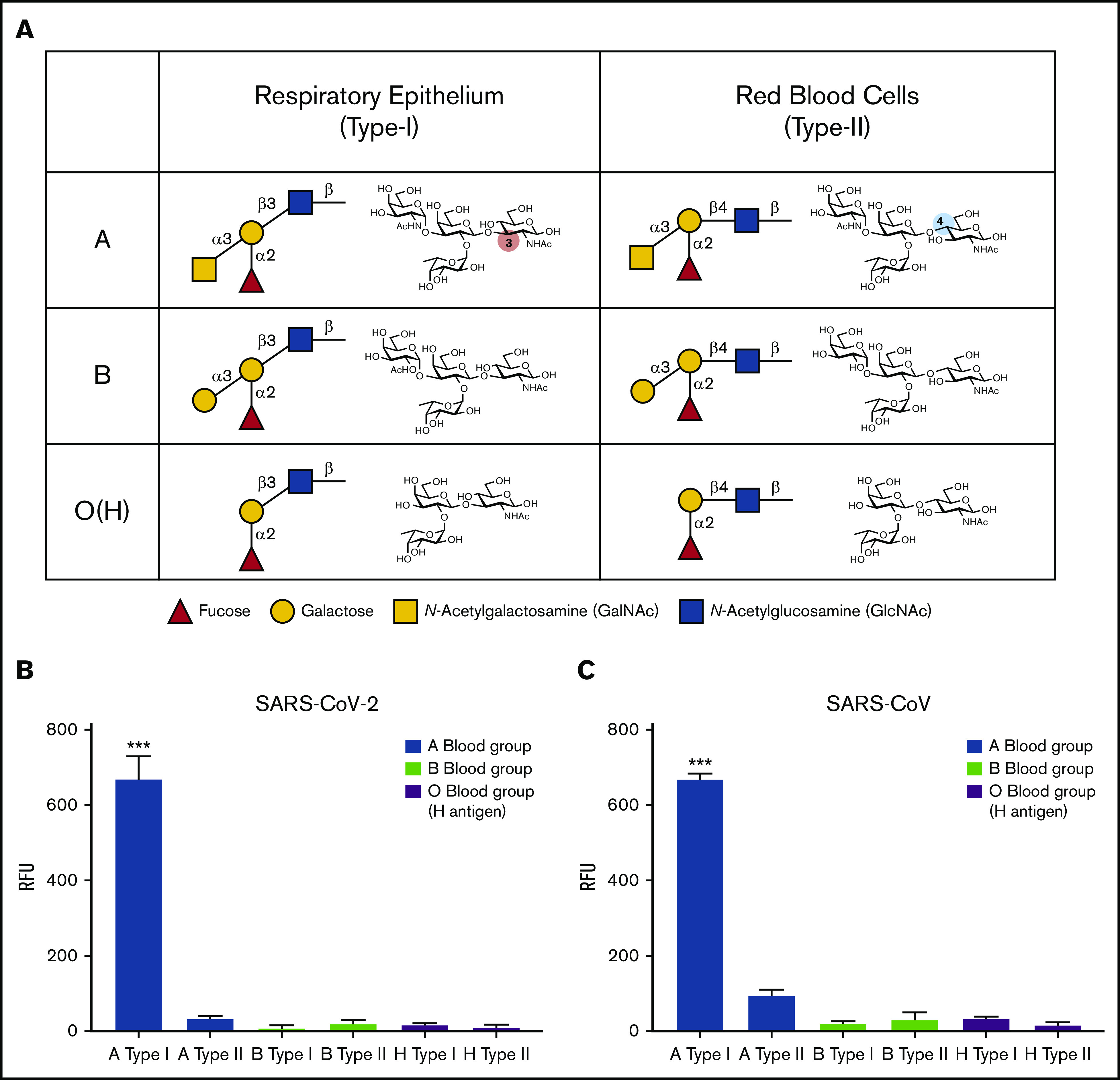
Figure 2.

SARS-CoV-2 RBD preferentially binds type I blood group A antigen. (A) Representation of human blood group antigens present on respiratory epithelium (type I) and RBCs (type II). Type I vs type II structures differ based on the linkage between the galactose and N-acetylglucosamine (type I: galactose β1-3 N-acetylglucosamine; type II: galactose β1-4 N-acetylglucosamine). (B-C) ABO(H) glycan microarray data obtained after incubation of SARS-CoV-2 RBD (B) or SARS-CoV RBD (C) with the corresponding glycans shown. Data are representative of 2 independent experiments. Error bars represent mean ± standard deviation. Statistics were generated by one-way analysis of variance with a post hoc Tukey’s multiple comparison. RFU, relative fluorescence unit. ***P < .001 for comparison between RBD binding to A type I vs all other glycans.
