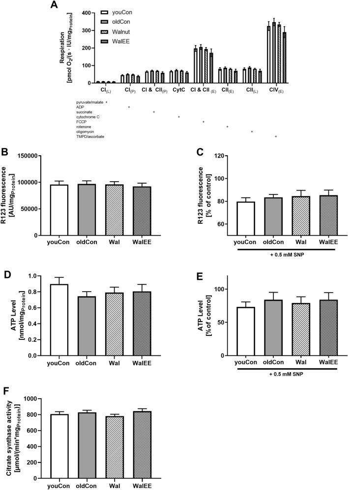
Fig. 3.
a Oxygen consumption of mitochondria isolated from the brain adjusted to protein. Activity of OXPHOS complexes were assessed via addition of several substrates, inhibitors or uncouplers. Which substance was added in which stage of the experiment is marked with “ + ”; n = 12. b Basal MMP levels were measured as R123 fluorescence after incubation of DBC samples. c MMP levels after 0.5 mM SNP-induced nitrosative stress. d Basal ATP concentration measured in µM per mg protein in the DBC samples. Measured signal stems from bioluminescence reaction of luciferin and ATP. e ATP levels after SNP-induced nitrosative stress. DBCs have been incubated for 3 h prior to measurement. f Citrate synthase activity as an indicator of mitochondrial content. Amount of protein was measured via BCA method. Displayed are means ± SEM. Statistical significance was tested via one-way ANOVA with Dunnett’s post hoc test using oldCon as reference for statistical comparisons. Each group in C-F consisted of 12–15 female NMRI mice. Parameters for the statistical tests can be found in the supplementary Table S2

