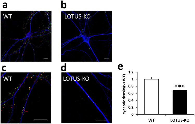
Figure 2.
Loss of LOTUS decreases the density of PSD95/Bassoon puncta in cultured hippocampal neurons. (a, b) Cultured hippocampal neurons (DIV 14) derived from WT (a) and LOTUS-KO (b) mice. Neurons were immunostained with antibodies against Bassoon (red), PSD-95 (green), and MAP2 (blue). Scale bars, 10 µm. (c, d) Magnified images from (a) and (b). The segment was imaged at 3 × magnification. Scale bars, 10 µm. (e) Quantification of the synaptic density of Bassoon/PSD95 puncta along the dendrites of each neuron. Data were normalized to the synaptic density in WT neurons. Data are means ± SEM from four to five independent experiments. The total number of neurons analyzed (n) ranged from 16 to 20 cells per condition. ***P < 0.001, Student’s unpaired t-test.

