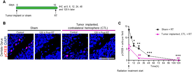
Figure 1.
Radiation induced acute damage of neuroblasts in the subgranular zone (SGZ). (A) intracranial GL261 tumor implantation or sham operation was performed in C57BL/6 mice followed by 10 Gy cranial irradiation (RT) at 10 dpi. Mice were euthanized at 0, 6, 12, 24, 48 and 120 h after RT and brain samples processed for IHC. (B) Representative images of pCREB immunostaining at 0 (control) and 120 h after RT in sham and tumor implanted mice. Images were
taken from the dentate gyrus (DG) of the hippocampus in the hemisphere contralateral (CTL) to the tumor implant site. Scale bar = 50 µm. (C) Quantification of pCREB positive cells in the SGZ at 0, 6, 12, 24, 48 and 120 h after RT. n = 3 per time point, *P < 0.05, **P < 0.01 and ***P < 0.001 compared to sham control and ##P < 0.01 compared to tumor implanted control mice by repeated ANOVA followed by Tukey’s post hoc t test.

