Figure 2.
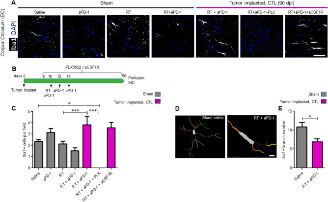
Tumor implanted mice treated with radiation in combination with aPD-1 showed chronic activation of microglia. (A) Sham operated mice were treated with either saline, aPD-1, RT or combined RT + aPD-1. Tumor implanted mice were treated with RT + aPD-1 plus either saline, PLX5622 or anti-CSF1R. Samples were collected at 90 dpi. Representative images of Iba1 IHC stain in the external capsule of the corpus callosum (EC) from the hemisphere contralateral to the implantation site (CTL) are shown. Scale bar = 50 µm. (B) Schematic of the experimental design for microglia elimination with PLX5622 and blockade of peripheral macrophages with anti-CSF1R blocking antibody in tumor implanted mice treated with RT + aPD-1. (C) Quantification of the numbers of Iba-1 cells in the EC of sham and tumor implanted mice. n = 6 per treatment arm in sham mice and n = 6 RT + aPD-1 + saline, n = 3 RT + aPD-1 + PLX5622 and n = 3 RT + aPD-1 + aCSF1R in tumor implanted mice, *P < 0.05, ***P < 0.001 by ANOVA followed by Tukey’s post hoc t test. (D) Fluorescent images of Iba1 + cells from sham mice and RT + aPD-1 treated tumor implanted mice were traced in Neurolucida software (https://www.mbfbioscience.com/neurolucida). Scale bar = 10 µm. (E) 3D reconstructions from the tracings were analyzed using the linear Sholl analysis method. The total number of branches per cell was significantly reduced in RT + aPD-1 treated tumor implanted mice. n = 3 per group, *P < 0.05 by Student’s t-test.

