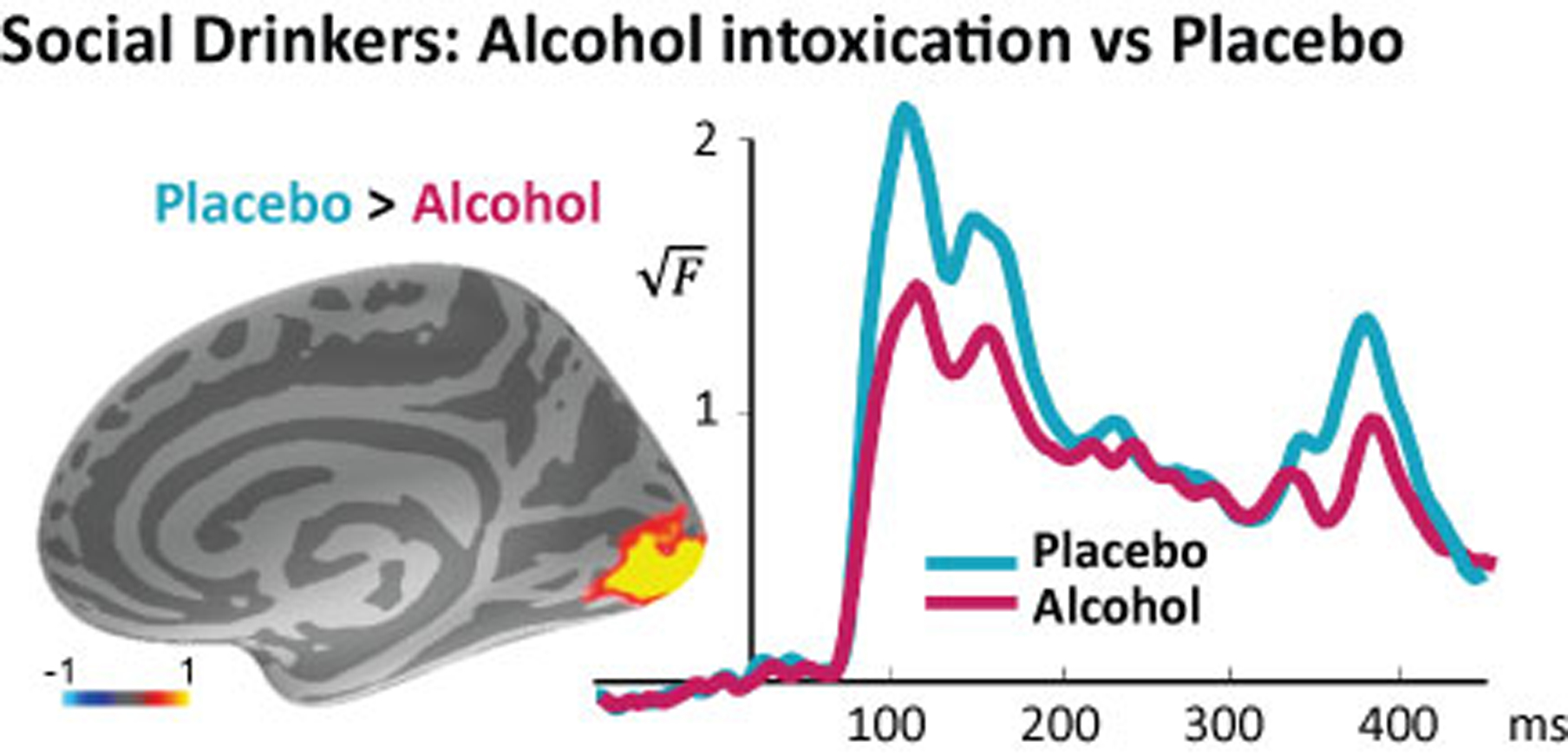
Figure 2.

MEG source estimate of the group average differential activity between placebo vs alcohol conditions (left) in the occipital cortex (Occ). Group average time courses of the estimated noise-normalized dipole strength in the Occ (right). Acute alcohol intoxication decreased the early visual activity in the occipital cortex within the time window indicated with a bar on the x-axis.
