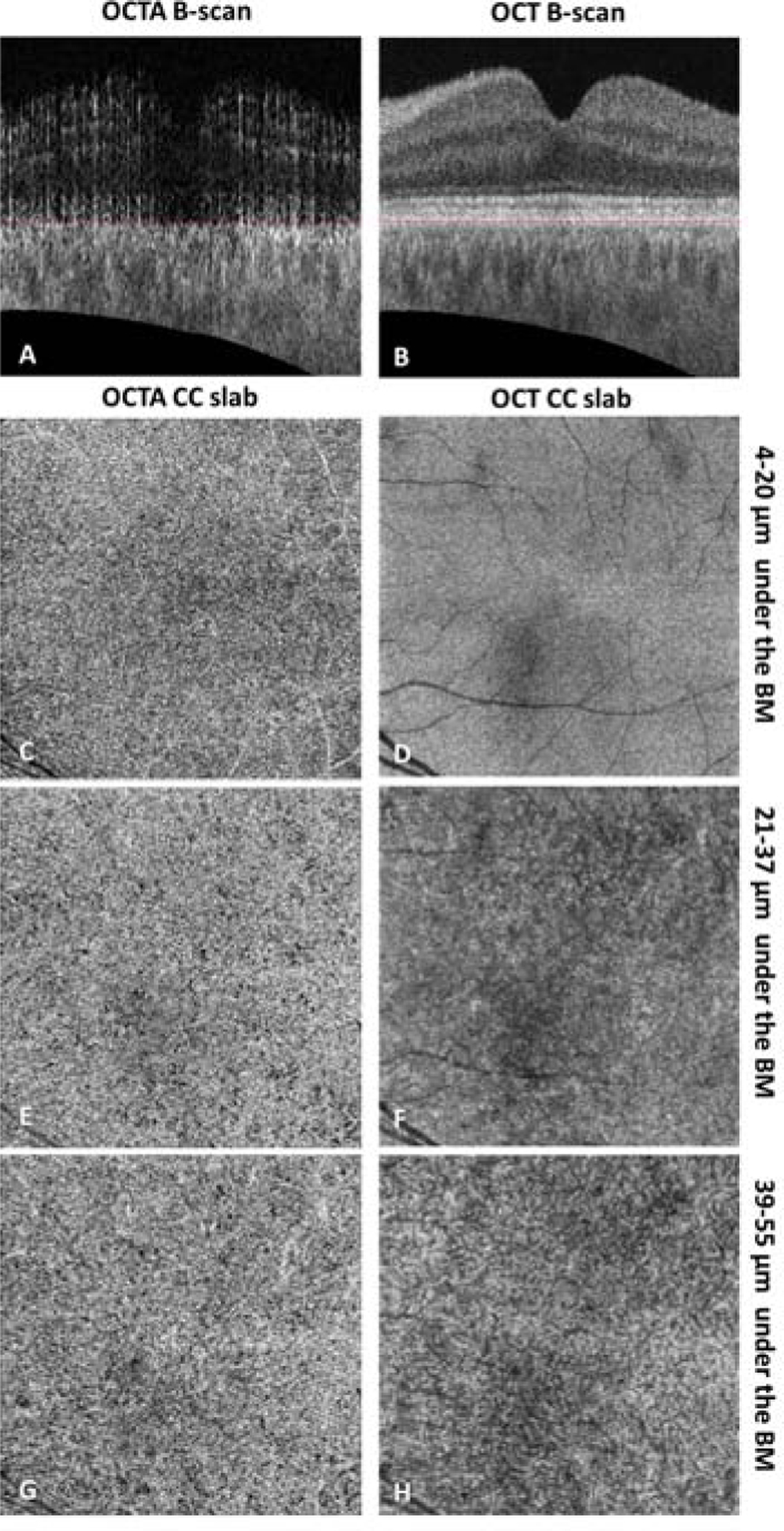
FIGURE 3.

Example of PLEX® Elite 9000 swept-source optical coherence tomography angiography (SS-OCTA) choriocapillaris (CC) slab with a thickness of 15 µm located at selected positions of a 6 × 6 mm scan in a normal eye. (A–B) Cross-sectional SS-OCTA and SS-OCT B-scans showing the position of segmented Bruch’s membrane (red dashed lines). (C–D) En face SS-OCTA and SS-OCT CC images with a position of 4–20 µm under the segmented BM. (Recommended) (E–F) En face SS-OCTA and SS-OCT CC images with a position of 21–37 µm under the segmented BM. (G–H) En face SS-OCTA and SS-OCT CC images with a position of 39–55 µm under the segmented BM. OCTA CC images have been compensated for shadowing effect by using the CC structural signals, and retinal projection artifacts have been removed. Positions in micron have been rounded (1 pixel = 1.9531 µm).
