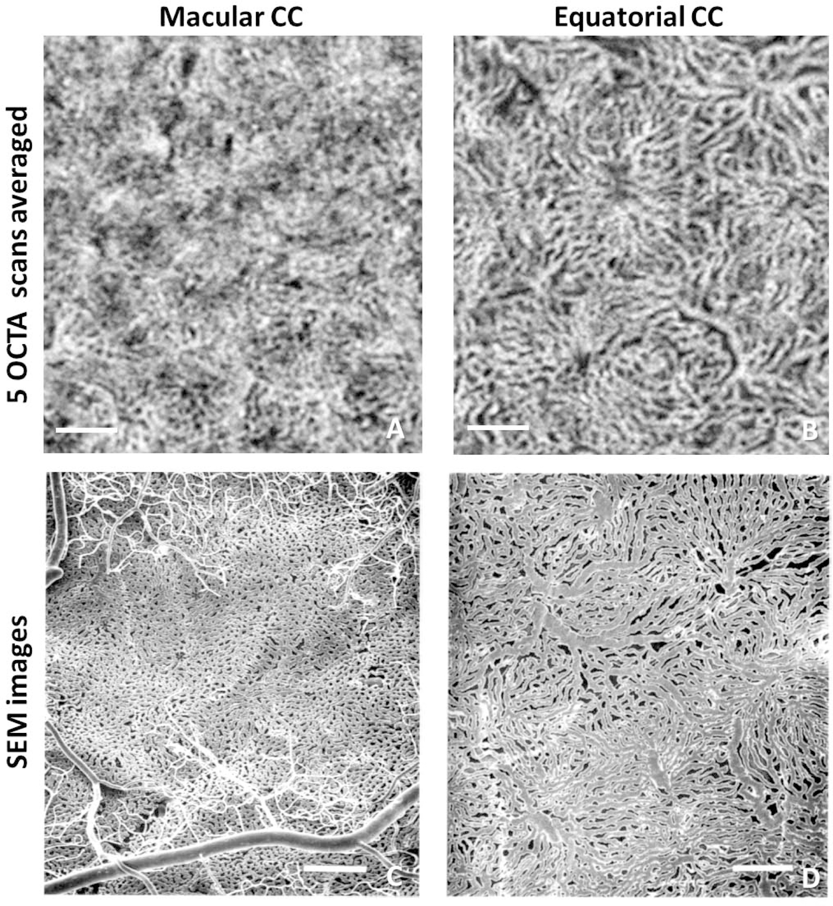
FIGURE 4.

Comparison of choriocapillaris (CC) in the macular region and equatorial region using PLEX® Elite 9000 swept-source optical coherence tomography angiography (SS-OCTA) and scanning electron microscopy (SEM). (A) En face SS-OCTA CC image acquired in the macular region, 5 3×3 mm scans averaged with 300 A-scans and 300 B-scan positions. (B) En face SS-OCTA CC image acquired in the equatorial region, 5 scans averaged (same as above). (C) SEM CC image of methyl methacrylate casts under the macular region reproduced from Olver et al. with permission.16 (D) SEM CC image of corrosion casts under the equatorial region reproduced from Olver et al. with permission.16 Scale bars: 250 µm. Reprinted by permission from Springer Nature: Springer Nature, EYE, Olver J. Functional anatomy of the choroidal circulation: methyl methacrylate casting of human choroid. Eye 1990;4:(2):262–272.
