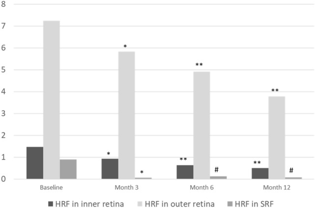
Figure 2.

The average number of HRF in different retinal layers at the baseline and 3, 6, 12 months after anti-vascular endothelial growth factor treatment for diabetic macular oedema. The number of HRF in every retinal layer significantly decreased in all follow up time points compared with baseline. HRF hyperreflective foci, SRF subretinal fluid. *p = 0.001 (compared with baseline). #p = 0.002 (compared with baseline). **p < 0.001 (compared with baseline).
